If you're looking for a dentist near me in Katy, TX because a tooth is missing, a bridge is failing, or you've finally decided to ask about dental implants near me, the first thing that usually surprises people is how much of the treatment starts before any surgery. The first step is almost always an image, because the team has to see what the eye can't. That picture helps answer the questions patients care about most, like whether the implant can fit safely, whether the bone is strong enough, and whether there's anything hidden under the gum that could change the plan.
At The Dental Retreat in Katy, that early imaging stage is handled as part of a calm, careful conversation, not a rushed checkbox. For patients in Sunterra, Cane Island, Katy Manor, Kingscrossing, Lakehouse, Marisol, The Grange, Anniston, Katy Lakes, Elyson, and Ventanna Lakes, that matters. The goal is simple, fewer surprises, clearer choices, and a treatment plan that feels understandable from the very beginning.
Why X-Rays Are the First Step in Any Implant Journey
A lot of people think implant treatment begins when the dentist places the post. It doesn't. It begins when the team looks at the jaw and decides whether the space, bone, and surrounding structures can support the new tooth safely.
The first look is about protection, not formality
A patient might come in after losing a tooth from decay, trauma, or a tooth extraction, and the instinct is to ask for a quick fix. The smarter move is to start with imaging, because hidden issues can change everything. Dental X-rays can reveal things that aren't visible during a regular exam, including bone damage, impacted teeth, abscesses, jaw abnormalities, and cavities that haven't shown up yet clinically, which is why they matter so much before restorative care begins (MedlinePlus dental X-ray overview).
That early picture is especially useful for implant planning because the team needs to know whether there's enough support before anything is placed. It also helps explain why a cosmetic dentist near me search often leads to the same answer as a restorative one, careful imaging comes first, then the treatment plan follows.
Practical rule: if the bone and surrounding anatomy haven't been mapped yet, no honest implant plan is complete.
A classic implant study analyzing 10,000 panoramic dental X-rays found that 5.43% of the radiographs showed at least one implant, totaling 1,791 implants, with an average of 3.2 implants per X-ray. It also found that only 1.22% of all implants showed X-ray findings consistent with lesions around the implant, which is one reason radiographic monitoring matters so much in implant care (study PDF).
Why patients usually feel less anxious after the first image
The first image changes the visit from guessing to planning. Patients often arrive worried about pain, cost, or whether they'll be told they need a bigger procedure than they expected. A good X-ray doesn't answer everything, but it does give the team a map to work from, and that alone can lower a lot of anxiety.
At a place like The Dental Retreat, that matters because implant care isn't just about technical accuracy, it's about helping people feel steady while decisions are being made. If you've been putting off an implant consult because you don't want surprises, the image is usually what turns the conversation into something concrete and manageable.
The Three Types of Dental Implant Imaging Explained

Think of implant imaging like choosing the right lens for a camera. A wide lens shows the whole scene, a close-up lens shows detail, and a 3D scan shows depth. Each one has a different job.
Panoramic X-rays give the broad map
A panoramic X-ray is the wide city map. It shows the full jaw in one sweep, which is useful for spotting general anatomy, existing teeth, overall bone patterns, and areas that may need more focused review. For implant planning, panoramic radiography is still described as the standard low-dose screening exam, and a PubMed review notes that it gives the best radiographic survey for treatment planning (PubMed review).
Periapical X-rays zoom in on one area
A periapical X-ray is the close-up of a single street corner. It focuses on a smaller region, so the dentist can inspect one tooth, one implant, or the bone around a specific spot. That close view is why it's so common in follow-up care, and why a 2020 survey of implantologists found that 84.3% routinely use periapical X-rays as the control radiograph (2020 survey PDF).
CBCT builds the 3D model
A CBCT scan, or cone beam CT, is the 3D drone footage. It shows height, width, depth, and bone density in a way a flat image can't. That's why it's often used when the team needs cross-sectional detail, especially for implant-site assessment and more complex surgical planning. Radiopaedia notes that CBCT provides 3D measurements of bone height, width, and density, and that the planned site typically needs at least 7 to 9 mm of height and 5 mm of width for implant placement (Radiopaedia dental implant article).
A flat X-ray can be enough for a straightforward check. A 3D scan becomes important when the dentist needs to see around corners.
When a Standard X-Ray Is Enough and When You Need a CBCT
Not every implant patient needs a 3D scan. That's the part many people never hear clearly, and it's where a lot of confusion starts. The right imaging tool depends on the question the dentist is trying to answer.
| Situation | Standard X-Ray | CBCT Scan |
|---|---|---|
| Routine implant follow-up | Often enough, especially with periapical imaging | Usually not needed for healthy, asymptomatic implants |
| Simple single-tooth implant planning | Often the first step | Used if bone depth, width, or anatomy needs cross-sectional detail |
| Suspected nerve or sinus issue | Can suggest a concern, but can't show full 3D relationship | Often needed to map the structure clearly |
| Complex anatomy or graft planning | May not give enough detail | Usually the better tool |
| Full-arch or All-on-4 planning | Limited for detailed spatial planning | Often preferred because angle and bone position matter more |
| Symptoms after placement | Helpful for a basic look | Used when there's a specific complication to investigate |
AAOMR guidance says not to use CBCT as the initial diagnostic exam and not for periodic review of asymptomatic implants, which is a more careful approach than many consumers expect (AAOMR position paper). That same practical idea shows up in patient-facing guidance from ADA and AAOMR materials, routine follow-up usually stays with conventional radiographs, while cross-sectional imaging is saved for presurgical planning, complications, or unusual anatomy (ADA-AAOMR patient selection PDF).
The easiest way to think about it is this, if the dentist just needs to check progress, a 2D image may be enough. If the dentist needs to map a jaw in three dimensions, the CBCT becomes the better tool. That difference matters for patients considering Emergency dentist care after an implant issue too, because symptoms often determine whether the next step is a quick X-ray or a deeper 3D look.
For readers asking about bone preparation, bone grafting for dental implants often comes into the conversation, because the scan tells the team whether the ridge needs help before placement.
How X-Ray Images Shape Your Implant Treatment Plan
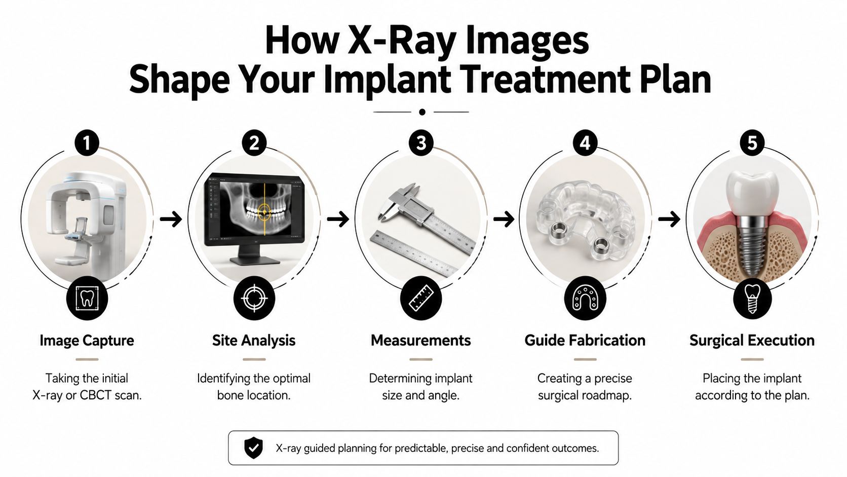
A scan matters when it changes what happens next. In implant dentistry, that means the image helps the team decide where the implant should sit, whether the site needs grafting, how long the implant should be, and how safely the procedure can be completed.
Bone space comes first
Before a dentist can talk about placement, the bone has to be checked for room. A standard X-ray gives a 2D view that can show overall bone height and nearby teeth, while a CBCT scan adds the side-to-side view that helps the team judge width and depth more accurately. That distinction matters because a ridge can look workable on a flat image and still be too thin in three dimensions.
Radiopaedia's dental implant article notes that the implant site typically needs at least 7 to 9 mm of height and 5 mm of width (Radiopaedia dental implant article). That is a practical guide for planning, not a guess. If the ridge is shorter or narrower than expected, the team may need to adjust the plan before surgery begins.
Edentulous ridges also tend to shrink in predictable ways, and the alveolar process often loses width before height. That matters because a site can look acceptable from the outside while still being too thin for a stable implant inside.
The image guides angle, depth, and safety
The scan also helps the dentist see where nerves, sinuses, and neighboring roots are located. A flat X-ray can be enough when the question is simple, such as checking whether a site appears clear for a straightforward implant. A CBCT scan becomes the better tool when the team needs to map the jaw in three dimensions, especially if the anatomy is tight, uneven, or close to sensitive structures.
That difference protects the patient from a placement angle that looks acceptable on the surface but crowds a structure underneath. In a single implant case, that may mean a small change in depth or angle. In a full-arch case, especially with All-on-4 planning, one poor angle can affect the whole arch, which is why cross-sectional imaging matters so much.
The Dental Retreat uses modern scanning and treatment planning technology for implant placement, and that matches the way implant planning works best, image first, then decide. For a plain-language look at the visit itself, see what to expect with a dental implant.
A scan becomes a surgical roadmap
Once the image is reviewed, the team can choose the implant size, compare placement options, and decide whether grafting should happen before or during placement. The scan also helps the dentist check whether a standard X-ray is enough or whether a CBCT is really needed for the case in front of them. That keeps the plan tied to the actual anatomy, instead of forcing every mouth into the same approach.
This is the value of imaging. It turns uncertainty into a step-by-step plan, and it lets the dentist explain the next move in plain language rather than guesswork.
A careful implant plan starts with measurements the team can trust.
What to Expect During Your Imaging Visit at The Dental Retreat
The imaging visit is usually much easier than patients expect. At The Dental Retreat on Stockdick School Road in Katy, the setting is designed to feel calm from the moment you walk in, with aromatherapy, heated massage chairs, noise-cancelling headphones, and in-room TVs helping the visit feel less clinical.
The appointment feels simple and controlled
For a panoramic X-ray, you'll stand or sit still while the machine moves around your head. For a CBCT scan, the process is similarly brief, and the goal is the same, capture the area clearly without asking you to do anything complicated. You may be asked to remove earrings, glasses, and dentures before the scan so the image stays clean and accurate.
Patients who feel anxious or claustrophobic usually do better when they know the room layout and the steps ahead of time. The staff can talk you through the positioning, help you stay comfortable, and make sure you understand what's happening before the scanner starts.
A few practical things help the visit go smoothly
- Bring your basics: a photo ID, insurance card if you have one, and any list of medicines you take.
- Leave metal accessories off: earrings, glasses, and removable appliances can interfere with imaging.
- Speak up early: if you've had a hard time with scans before, tell the team before you sit down.
- Ask questions before you leave: the image is only useful if you understand what the dentist saw.
The biggest relief for many patients is realizing there's no pressure to “perform” during the scan. You just stay still while the team does the technical work. That's especially reassuring for people coming in for new patient exams, dental implants, or a dentist in Katy, TX visit after months of putting treatment off.
Understanding Radiation Safety in Dental Implant Imaging
Many patients worry more about the radiation than the scan itself. That fear is understandable. Few individuals receive a clear explanation, so the word “radiation” sounds bigger and scarier than it usually is in dental care.
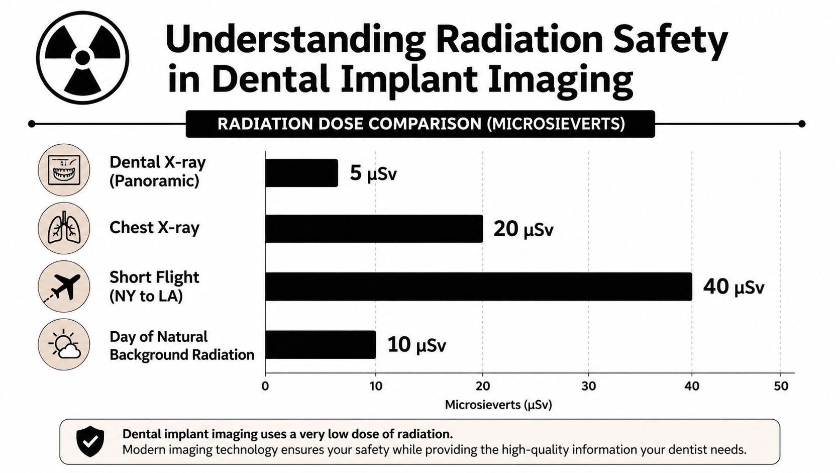
The dose is managed carefully, not casually
Dental imaging follows the ALARA principle, which means the team uses as little radiation as reasonably possible while still getting the diagnostic information needed. One practical way that happens is by using the smallest field of view that still captures the surgical site, because a narrower field reduces scatter and unnecessary exposure.
The service guidance on imaging notes that a single-implant CBCT benchmark is a 4 cm diameter by 4 cm height cylindrical volume, and that published guidance cites an achievable adult dose around 250 mGy·cm² for that procedure (HPA-RPD-065 PDF). It also says intraoral units operate optimally around 60 to 70 kVp, while settings above 90 kVp increase patient dose. That's why a well-run practice doesn't just take any image, it chooses settings and field size with care.
Routine questions come up for good reason
Patients often ask whether repeated imaging adds up. The answer is that the team doesn't order images by habit, it orders them when the result changes the plan. That's the whole point of dose control, enough information to treat safely, not more exposure than the case needs.
Radiation safety in implant imaging is mostly about discipline, choosing the right scan, the right size, and the right timing.
The earlier comparison between panoramic, periapical, and CBCT imaging helps here too. If a 2D image can answer the question, there's no reason to jump straight to a 3D scan. That restraint is part of good care, whether the patient is seeking restorative dentistry, tooth extraction, or implant planning after missing a tooth.
Long-Term Monitoring with X-Rays After Your Implant Is Placed
Imaging does not stop once the implant is placed. Many patients assume the hard part is over after surgery, but follow-up X-rays are part of the full implant journey because the bone and surrounding tissues still need time, and they still need attention. A healthy implant is not watched once and forgotten. It is checked over time, the same way a clinician keeps following a healing fracture or a new restoration to see whether the support stays stable.
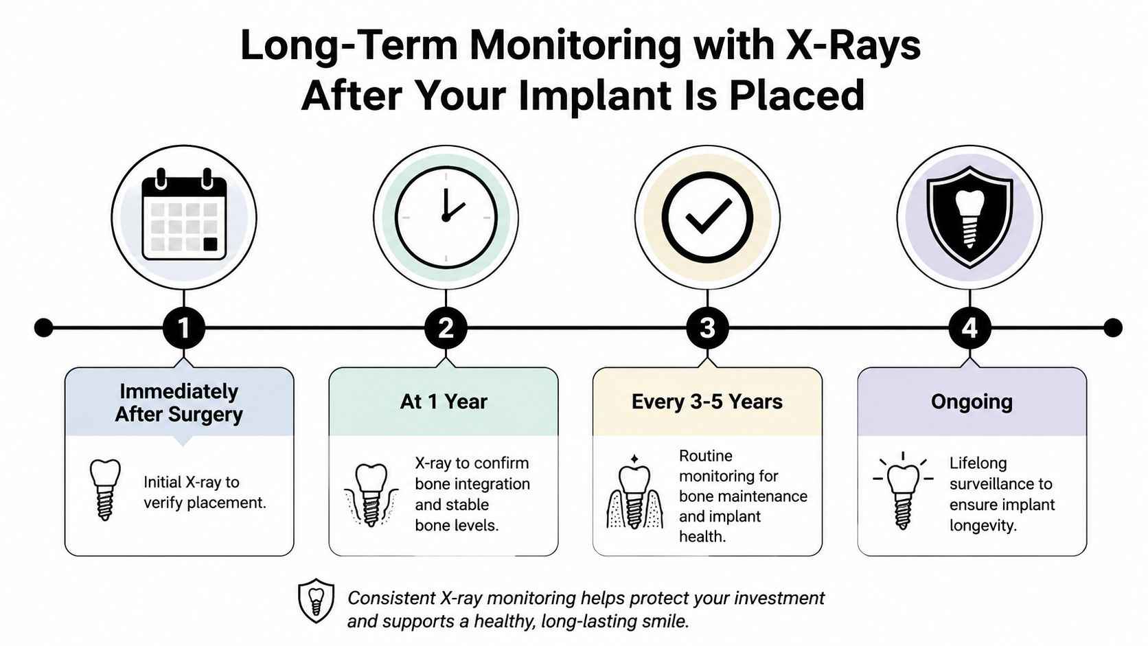
Follow-up X-rays check the details that matter
A PubMed implant imaging article explains that radiographs are needed before treatment, useful after healing to verify the bone next to the implant and to check that transmucosal abutments fit properly, and also appropriate after the prosthesis is completed so the team can watch both early and long-term results (PubMed implant imaging article). That is the rhythm of implant care. The image at the start helps set the plan, and later images show whether the plan is holding up.
A study PDF on implant outcomes also reported radiographic findings around implants at a low rate, which supports the idea that problems are not expected, but they are still worth checking for when care is being maintained (study PDF). A quiet result on an X-ray does not mean the implant can be ignored. It means the team has a baseline to compare against if anything changes later.
A good X-ray gives information, not a promise
A follow-up X-ray can show stable bone levels and help the team notice small changes before they become obvious in the mouth. It cannot promise the implant will last forever. It works more like a regular status check on a repaired bridge, one snapshot showing whether the support still looks steady.
That is why routine maintenance often stays with conventional radiographs, while CBCT is reserved for symptoms, complications, or a specific diagnostic question (ADA-AAOMR patient selection PDF). A plain X-ray is often enough when the question is simple, such as whether bone levels look stable or whether the crown and abutment area look as expected. A CBCT scan is used when the dentist needs a three-dimensional view, usually because the regular film does not answer the question clearly enough. For anxious patients, that difference can make the process feel less mysterious, because imaging becomes a careful long-term check rather than a sign that something has already gone wrong.
Common Questions Katy Patients Ask About Implant X-Rays
A lot of the same questions come up in every consult, and they deserve straight answers. Patients want to know what the first visit looks like, whether the scan is included, and how the plan changes if they need a full-arch solution instead of one implant.
What happens at a new patient exam
A visit usually starts with a conversation, a clinical exam, and imaging if it's needed to understand the problem. For patients without insurance, The Dental Retreat also offers a $99 exam with cleaning and X-rays, along with a $49 problem-focused visit and $350 in-office Zoom whitening, which can make it easier to get started without guessing about cost. That matters for people who have been delaying care and now need clear next steps.
How the team decides what imaging you need
The decision is based on the question, not the menu of machines. A routine follow-up might only need a periapical film, a single-tooth implant may begin with a panoramic X-ray, and a more complex case may call for CBCT because the dentist needs depth and cross-sectional detail. That same logic applies whether you're asking about a single implant, All-on-4, or a broader emergency dentist concern after pain or swelling.
Which neighborhoods this matters for
People in Katy, TX, and nearby communities like Sunterra, Cane Island, Katy Manor, Kingscrossing, Lakehouse, Marisol, The Grange, Anniston, Katy Lakes, Elyson, and Ventanna Lakes often want one simple answer. Do I need another scan, or is the standard X-ray enough? The honest answer is that the right image depends on the stage of care, and a good implant team should be able to explain that in plain English before anything is scheduled.
If you're ready to find out what your jaw needs, schedule a visit with The Dental Retreat. Their team can walk you through the imaging, explain whether a standard X-ray or CBCT makes sense for your case, and help you move forward with a clear plan that fits your comfort and your goals.